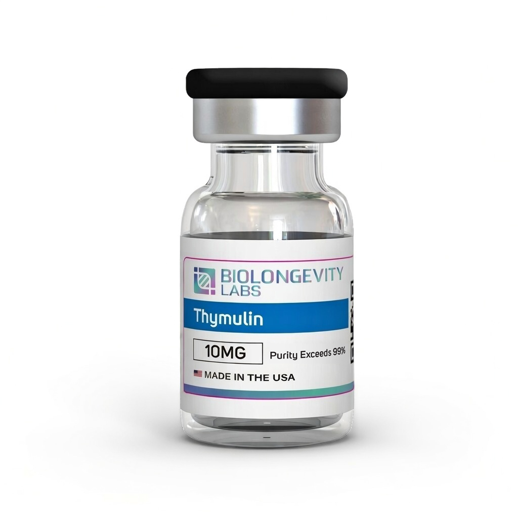
Thymulin peptide for investigating thymic hormone mechanisms in immune cell differentiation. High-purity lyophilized powder for cellular research. Research use only compound.
Thymulin is a nonapeptide hormone (nine amino acids) originally isolated from thymus tissue, studied for its role in immune system regulation and T-cell differentiation mechanisms. This research-grade compound is supplied as lyophilized powder with high purity standards verified through advanced analytical testing.
The molecular structure (Pyr-Ala-Lys-Ser-Gln-Gly-Gly-Ser-Asn-OH) enables investigation of thymic factor activity in immune cell development and cellular signaling pathways. Each batch undergoes rigorous chemical characterization to ensure consistent quality for laboratory applications.
Manufactured in USA GMP facilities with comprehensive documentation. This peptide compound is intended exclusively for in vitro research investigating immune function mechanisms.
Thymulin is a nonapeptide (pyro-Glu-Ala-Lys-Ser-Gln-Gly-Gly-Ser-Asn) originally identified as facteur thymique sérique (FTS).
Molecular Identity and Zinc Dependence
The peptide requires zinc binding in a 1:1 ratio to achieve cellular activity.
NMR studies confirmed that zinc binding induces a specific three-dimensional conformation required for receptor binding. Thymic epithelial cells secrete the peptide in its active zinc-containing form.[1]
Zinc deficiency models show that reduced thymulin activity directly correlates with declines in T-cell subpopulations and lymphokine production. Zinc repletion consistently restores thymulin bioactivity in laboratory investigations.[2]
Immune Modulation
T-Cell Differentiation and Th1/Th2 Balance
Thymulin influences both intrathymic and extrathymic T-cell differentiation through specific receptor binding on T-cell lines.
Gene expression studies in murine asthma models demonstrated thymulin’s ability to shift immune balance away from Th2 dominance:[3]
Increased IFN-γ expression, stimulating Th1 cells
Reduced IL-13 secretion (Th2-associated mediator)
Decreased eotaxin (eosinophil chemotactic factor)
Lower eosinophil, neutrophil, and mononuclear cell infiltration in bronchoalveolar lavage fluid
Zinc deficiency investigations show that reduced thymulin activity impairs Th1 cell proliferation while leaving Th2 function intact, resulting in measurable Th1/Th2 imbalance.[4]
Anti-Inflammatory Mechanisms
Cytokine and Mediator Downregulation
A thymulin peptide analogue (PAT) demonstrated anti-inflammatory activity in endotoxin-induced inflammation models, downregulating multiple proinflammatory mediators simultaneously.[5]
Mediator
Effect of PAT
Model
TNF-α
Abolished elevation
Localized (i.pl. ET)
IL-1β
Reduced levels
Localized and systemic
IL-6
Reduced to control levels
Localized and systemic
NGF
Reduced levels
Localized (i.pl. ET)
PGE₂
Reduced to control levels
Systemic (i.p. ET)
Mechanistic data indicate PAT inhibits NF-κB nuclear translocation, the transcription factor governing proinflammatory cytokine and COX-2 gene expression.
Separate mouse models showed thymulin prevented overproduction of proinflammatory cytokines and heat shock protein Hsp70 through the NF-κB pathway.[6]
Anti-Fibrotic Signaling
Thymulin gene expression in asthma models reduced airway remodeling markers:[3]
Decreased TGF-β and VEGF expression
Reduced collagen fiber deposition in airways
Attenuated smooth muscle hypertrophy in terminal bronchioles
Prevented subepithelial fibrosis and mucous cell hyperplasia
Earlier work showed thymulin reduced bleomycin-induced cytokine production and inhibited pulmonary fibrosis. In monocrotaline-induced pulmonary hypertension, thymulin suppressed the proinflammatory p38 MAPK pathway.
Neurological Research
Analgesic Properties
PAT exhibited analgesic potency comparable to dexamethasone and indomethacin in reversing endotoxin-induced mechanical and thermal hyperalgesia, but at lower molar concentrations. It outperformed anti-IL-1β peptides by approximately 10x in molar terms.[5]
PAT reversed both hyperalgesia and fever in systemic inflammation models. Effects are attributed to suppression of COX-2-dependent PGE₂ formation and peripheral cytokine cascades.
Neuroprotection
Intracerebroventricular administration studies demonstrated thymulin’s neuroprotective role in experimentally induced brain inflammation, reversing inflammatory hyperalgesia and modulating proinflammatory cytokine concentrations.[7]
Adenoviral vectors carrying synthetic thymulin genes achieved longer transgene expression in rat brain regions compared to other reporter genes. The proposed mechanism involves thymulin’s local anti-inflammatory activity protecting virus-transduced brain cells from immune-mediated destruction.
Neuroendocrine Regulation
Hypophysiotropic Activity
Thymulin acts as a hypophysiotropic peptide, regulating pituitary hormone release in dispersed rat pituitary cell studies:[7]
Luteinizing hormone (LH) and follicle-stimulating hormone (FSH) release
Growth hormone (GH) and prolactin (PRL) secretion
Thyrotropin (TSH) release
Adrenocorticotropic hormone (ACTH) secretion via cAMP and cGMP accumulation
Thymulin also modulates GnRH stimulatory activity on gonadotropin release and influences gonadotropin-induced ovarian and testicular steroidogenesis.
Bidirectional Regulation
Thymulin production is regulated by the neuroendocrine system in a bidirectional loop. Growth hormone stimulates thymulin release from thymic epithelial cells via GH receptors.
Prolactin receptors on thymic epithelial cells allow direct stimulation of thymulin synthesis. Thyroid hormones (T₃/T₄) stimulate thymulin secretion, while hypothyroidism depresses circulating levels.
Metabolic Research
Athymic mouse models develop spontaneous hyperglycemia, impaired glucose tolerance, and peripheral insulin insensitivity after approximately one month. These mice show increased somatostatin-producing D-cell populations and elevated pancreatic somatostatin content.[7]
Partial normalization of hepatic phospholipid fatty acid composition
These findings indicate thymulin’s modulatory role in both glucose and lipid metabolism pathways.
Aging and Thymic Involution
Age-related thymus involution results in progressive decline of thymulin secretion, contributing to immunosenescence in aging organisms. Thymic involution has been linked to cellular senescence driven by oxidative stress.[8]
Several neuroendocrine interventions demonstrated partial restoration of declining thymulin levels in aged animal models. Bovine GH treatment in aged dogs partially restored low serum thymulin levels.
Ovine GH in old mice increased circulating thymulin and enhanced thymocyte proliferation and IL-6 production. Combined GH and T₄ treatment partially restored thymulin levels in old rats.
Extrathymic Production Under Cellular Stress
Studies show that thymulin is not exclusively of thymic origin. When non-thymic cells (RAW 264.7 macrophages and L929 fibroblasts) were exposed to oxidative stress, heat shock, or apoptosis-inducing agents, extracellular thymulin was detected in culture media within 2 hours.[6]
SPATS2L as Putative Precursor
Western blot analysis revealed two bands (~60 kDa and ~10 kDa) interpreted as potential precursor and intermediate product. SPATS2L (Spermatogenesis-Associated Serine-Rich 2-Like), an intranucleolar stress-response protein, was identified as containing a thymulin-like sequence.
Key observations supporting SPATS2L as a thymulin precursor:
Strong Pearson correlation (r = 0.925, p < 0.01) between SPATS2L and 60 kDa thymulin-stained band
SPATS2L migrates from nucleolus to stress granules under oxidative stress
The 10 kDa intermediate correlates with caspase-3 activation
The 60 kDa precursor correlates with NF-κB pathway activation
This extrathymic production pathway suggests thymulin may function as a stress-responsive signal from damaged cells to local immune cells, potentially explaining how thymulin-mediated immune regulation persists after age-related thymic involution.
Summary of Signaling Pathways
Pathway / Target
Effect of Thymulin
Study Context
NF-κB
Inhibits nuclear translocation
LPS inflammation models
p38 MAPK
Suppresses activation
Pulmonary hypertension
Th1/Th2 balance
Shifts toward Th1 (↑IFN-γ)
Asthma, zinc deficiency
COX-2 / PGE₂
Reduces PGE₂ production
Systemic inflammation
TGF-β / VEGF
Downregulates expression
Airway remodeling
cAMP / cGMP
Mediates ACTH stimulation
Pituitary cells
HMG-CoA reductase
Inhibits activity
Cholesterol metabolism
Research Use Only: This compound is intended exclusively for in vitro laboratory research.
References
Dardenne M, Pléau JM, Nabarra B, Lefrancier P, Derrien M, Choay J, et al. Contribution of zinc and other metals to the biological activity of the serum thymic factor. Proceedings of the National Academy of Sciences; 1982. https://doi.org/10.1073/pnas.79.17.5370
Prasad AS, Meftah S, Abdallah J, Kaplan J, Brewer GJ, Bach JF, et al. Serum thymulin in human zinc deficiency. American Society for Clinical Investigation; 1988. https://doi.org/10.1172/jci113717
da Silva AL, Martini SV, Abreu SC, Samary C dos S, Diaz BL, Fernezlian S, et al. DNA nanoparticle-mediated thymulin gene therapy prevents airway remodeling in experimental allergic asthma. Elsevier BV; 2014. https://doi.org/10.1016/j.jconrel.2014.02.010
Safieh‐Garabedian B, Dardenne M, Pléau JM, Saadé NE. Potent analgesic and anti‐inflammatory actions of a novel thymulin‐related peptide in the rat. Wiley; 2002. https://doi.org/10.1038/sj.bjp.0704793
Lunin SM, Khrenov MO, Glushkova OV, Vinogradova EV, Yashin VA, Fesenko EE, et al. Extrathymic production of thymulin induced by oxidative stress, heat shock, apoptosis, or necrosis. SAGE Publications; 2017. https://doi.org/10.1177/0394632017694625
Barbouti A, Vasileiou PVS, Evangelou K, Vlasis KG, Papoudou-Bai A, Gorgoulis VG, et al. Implications of Oxidative Stress and Cellular Senescence in Age-Related Thymus Involution. Wiley; 2020. https://doi.org/10.1155/2020/7986071
Certificate of Analysis (COA) for Every Batch
A Certificate of Analysis (COA) is a document that verifies a compound’s identity, purity, and batch quality through independent laboratory testing. Every compound from BioLongevity Labs ships with a COA tied to its specific batch, so researchers can confirm exactly what they received before it enters a protocol.
Each COA reports results from third-party laboratory analysis, including:
High-performance liquid chromatography (HPLC) for purity, typically confirmed at 99% or higher
Liquid chromatography mass spectrometry (LC-MS) for molecular identity and mass confirmation
Sterility and endotoxin screening where applicable
Chemical contaminant and residual solvent checks
COAs are sourced from independent certified labs rather than in-house testing alone, giving researchers a verifiable record of molecular integrity for each batch. All compounds are supplied for research use only.
Scientifically reviewed by Dr. Ky H. Le, MD. Dr. Le is a board-certified family medicine physician with over 20 years of clinical experience. Dr. Le validates the scientific accuracy of all technical content and research citations.
Disclaimer: For Research Purposes Only
This content is provided strictly for research purposes and does not constitute an endorsement or recommendation for the non-laboratory application or improper handling of peptides designed for research. The information, including discussions about specific peptides and their researched benefits, is presented for informational purposes only and must not be construed as health, clinical, or legal guidance, nor an encouragement for non-research use. Peptides described here are solely for use in structured scientific study by authorized individuals. We advise consulting with research experts, medical practitioners, or legal counsel prior to any decisions about obtaining or utilizing these peptides. The expectation of responsible, ethical utilization of this information for legitimate investigative and scholarly objectives is paramount. This notice is dynamic and governs all provided content on research peptides.